Blood Collection Tubes to Collecting Whole Blood

Blood Collection Tubes
Blood collection tubes are sterile, single-use vacuum tubes designed to safely collect venous blood for laboratory tests. Each tube contains a predefined vacuum and, if needed, a specific additive (anticoagulant, clot activator, preservative, or separator gel) tailored to the type of test. The vacuum passively draws blood into the tube once the patient’s vein is accessed, making collection safer and more convenient. Tubes are color-coded by cap shank (per ISO 6710) to indicate the additive inside. For example, light-blue tops hold sodium citrate (an anticoagulant for coagulation studies), lavender (purple) tops hold EDTA (a calcium-chelator for hematology), green tops hold heparin, and red or gold tops have no additive (or a clot activator plus gel) for serum chemistry tests. In short, the cap color “tells you exactly what additive is inside, and the additive determines what happens to the blood after collection”.
Blood collection tubes (often called vacutainer tubes) are sterile glass or plastic tubes pre-filled with a vacuum. When venipuncture is performed, the vacuum draws a precise volume of blood into the tube. Each tube has a color-coded cap indicating which additive (if any) is inside. These additives (anticoagulants, clot activators, separating gels, or preservatives) stabilize the blood for specific laboratory tests. For example, anticoagulants like EDTA, citrate, or heparin prevent clotting so that plasma or whole blood can be tested; clot activators (silica particles) induce clotting to yield serum; and gels (in “separator” tubes) form a barrier between cells and serum after centrifugation Some tubes also contain preservatives (e.g. sodium fluoride) that inhibit metabolic reactions (such as glycolysis) to stabilize analytes like glucose.
Types of Tubes (Color and Additive)
Common blood collection tubes (with their typical additives and uses) include:
-
Light Blue Top (Citrate): Contains 3.2% sodium citrate anticoagulant. Used for coagulation studies (e.g. prothrombin time PT and activated partial thromboplastin time aPTT). Citrate binds calcium to prevent clotting, producing plasma upon centrifugation. Because accurate testing requires a strictly 9:1 blood-to-citrate ratio, the tube must be filled to the marked line. After draw, immediately invert gently to mix.
-
Red Top (Plain/Serum): No anticoagulant (glass tube) or contains clot activator (plastic tube). Blood in a red-top tube clots naturally; after 10–60 minutes at room temperature the clot is removed by centrifugation to yield serum. Red tops are used for general chemistry and serology tests that require serum. (Note: For many chemistries, a “tiger-top” or Gold SST tube is preferred.)
-
Gold or “Tiger-Top” (SST – Serum Separator Tube): These have a clot activator plus an inert gel. The gel forms a barrier between clot and serum when centrifuged. They are colloquially called gold, marble-top, or tiger-top tubes. Use gold SST tubes for chemistry panels, immunoassays, and other tests on serum. After collection, invert 5–10 times to mix clot activator, allow clotting (≈30–60 min), then centrifuge. (CDC recommends waiting at least 30 min up to 60 min for complete clot formation.)
-
Green Top (Heparin): Coated with heparin (either sodium heparin or lithium heparin). Heparin inhibits thrombin, preventing clotting so that plasma is obtained. Green-top tubes are used for many STAT plasma chemistries (e.g. electrolytes, chemistry panels, blood gases). There is also a light-green/Lithium Heparin tube with a gel separator (PST) for plasma—used similarly in chemistry.
-
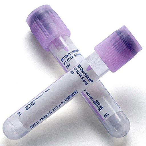
Lavender (or Purple) Top (EDTA): Contains K₂EDTA anticoagulant. EDTA chelates calcium and prevents clotting. Lavender top tubes yield whole blood or plasma and are the standard for hematology (e.g. CBC) and blood bank compatibility testing. Pink-top tubes are similar (K₂EDTA) and are typically used for transfusion crossmatch and other immunohematology tests.
-
Gray Top (Oxalate/Fluoride): Contains potassium oxalate (anticoagulant) and sodium fluoride (glycolysis inhibitor). Used for glucose and lactate measurements (fluoride stabilizes blood sugar by inhibiting enzymes) and for alcohol levels. After draw, invert to mix additives.
-
Yellow Top (ACD or SPS):
- ACD (Acid-Citrate-Dextrose): Some yellow-top tubes contain ACD solution, used for specialized tests (e.g. HLA phenotyping, DNA/PBMC collection, autosomal genetic tests) because it preserves cells.
- SPS (Sodium Polyanethol Sulfonate): Blood culture bottles (for microbiology) often have yellow or pink stoppers. These contain SPS, which prevents clotting and inhibits complement. (Don’t confuse these culture bottles with the serum/plasma tubes above.)
-
Other Colors: Specialty tubes include royal blue (trace element tubes, either EDTA or clot-activated, for lead/analyses), tan (lead EDTA), black (citrate for erythrocyte sedimentation rate), orange (rapid serum tubes with thrombin), and others. These are used only for specific tests. The exact coloring and additives are standardized (CLSI) but can vary slightly by manufacturer
Venipuncture Technique and Order of Draw
Patient Preparation: Always use two patient identifiers (e.g. name and DOB) and confirm them against the lab requisition before drawing blood. Obtain consent and explain the procedure. Address any latex sensitivities (use non-latex tourniquets and gloves if needed).
Equipment: Use a sterile needle (typically 21–23 gauge for adults; 23–25 G butterfly for children or difficult veins) with a holder (vacutainer adapter) and the required tubes. Check each tube for an intact seal and suitable expiration date; a tube with a compromised vacuum will not fill properly.
Vein Selection: Apply the tourniquet 7–10 cm above the intended puncture site to engorge veins, but no longer than 1 minute. Palpate veins (usually median cubital in the antecubital fossa). Clean the site with 70% isopropyl alcohol and allow it to air-dry (~30 seconds). Do not touch the site after disinfection.
Order of Draw: If multiple tubes are needed, follow the CLSI-recommended sequence to prevent additive carry-over:
- Blood cultures (sterile bottles) – first, to avoid contamination.
- Coagulation tube (citrate, light blue) – next, to avoid contamination by EDTA or clot activators. (Always fill citrate tubes to the fill line for a 9:1 ratio.)
- Serum tubes (red or gold SST) – for chemistry/serology.
- Heparin tubes (green) – for stat chemistries.
- EDTA tubes (lavender or pink) – for hematology/blood bank.
- Glycolytic inhibitor tubes (gray) – last, as additives here can alter other tests.
Follow any institutional variations, but the principle is “sterile first, then citrate, then serum, then heparin, then EDTA, then gray”. Always gently invert each tube immediately after collection to mix blood with additive (see below) and label each tube at the bedside with patient ID, date/time, and initials.
Drawing the Blood: Insert the needle with the bevel up at a shallow angle (~15°) through the skin into the vein. Stabilize the needle and holder arm with your hand. Once blood flow begins, release the tourniquet as soon as enough blood is flowing (to avoid hemoconcentration. Lower the patient’s arm (to ensure flow) and allow each tube to fill under vacuum. Do not remove the needle or the tube during filling; the vacuum will automatically draw the correct volume if the tube seal and vacuum are intact.
After the Draw: Remove the last tube, then the needle. Release the tourniquet before removing the needle to prevent hematoma. Immediately cover the puncture site with clean gauze and ask the patient to apply pressure until bleeding stops (especially important if on anticoagulants). Dispose of the needle in a sharps container (no recapping) and place used tubes in a biohazard rack or container for transport. Label all tubes securely before leaving the patient.
Mixing and Handling of Samples
Right after collection, mix anticoagulated tubes by gently inverting them end-over-end 5–10 times (as per manufacturer). A full inversion is 180° to 0° and back. This ensures the blood thoroughly contacts the anticoagulant or clot activator. Do not shake the tubes vigorously, as that can cause hemolysis or microclots. For example, CLSI guidelines state that inadequate mixing leads to clotting or hemolysis, producing inaccurate results. Once mixed, keep the tubes upright.
Clotting (for serum tubes): After drawing blood into a clot-activator tube (red/gold), allow it to clot at room temperature (typically 15–30 minutes) before centrifugation. Do not centrifuge wet clots – fibrin strands can contaminate serum and clog analyzers. After clotting, centrifuge as soon as possible (ideally within 2 hours). Serum/plasma separated from cells promptly minimizes analyte changes (cells metabolize glucose, etc.).
Transportation: Carry tubes to the lab immediately in a tube rack or sealed transport box to avoid breakage or spillage. If tests are delayed, follow handling instructions (some require refrigeration, light protection, etc.). For example, some specimens (like ammonia) require chilling on ice. Label each sample clearly and ensure documentation matches.
Collection and Handling
Blood is drawn using a double-ended Vacutainer needle and holder system. After venipuncture, the first tube is pushed onto the holder’s inner needle; the tube cap is punctured and its vacuum pulls blood into the tube. When the tube fills, it is removed and the next tube is inserted. Each tube’s vacuum stops drawing once its preset volume is reached.
Key handling steps:
- Mixing: Immediately after filling, tubes containing anticoagulant must be gently inverted (usually 5–10 times) to mix blood with the additive. Overly vigorous shaking can cause hemolysis; under-mixing can allow small clots to form.
- Clot formation: Serum tubes (red, gold) require time to clot before centrifugation (~30–60 min).
- Labeling: Tubes should be labeled at the bedside with patient ID and collection time before leaving the patient to avoid misidentification.
- Storage: After collection, tubes are transported to the lab (some tests require prompt processing or specific temperature).
All tubes are single-use and sterile. They bear manufacturer expiration dates: an expired tube may have lost its vacuum and will not fill properly. Always check expiry before use.
Indications and Tests
Doctors and nurses choose tubes based on the ordered tests. Key associations are: a lavender-top EDTA tube is used for CBC/Hematology; a light-blue citrate tube for coagulation profiles (e.g. PT/INR); red or gold-top serum tubes for general chemistry and immunology panels); a green-top heparin tube for stat chemistries and some hematology DNA tests; and a gray-top tube for glucose and alcohol assays. For example, drawing a potassium level in an EDTA tube will artifactually raise potassium (EDTA releases intracellular potassium), and drawing a coagulation test in a plain tube will allow clotting before analysis. Thus, using the correct tube is essential for valid results.
Tips, Precautions, and Best Practices
- Patient Identification: Always re-check patient ID (name/DOB) with the armband and lab request to prevent mix-ups. Mislabeling is a serious error.
- Sterile Technique: Never touch the tip of the needle, and keep the puncture site from recontamination after cleaning. Use gloves and change gloves if they become contaminated.
- Tourniquet Use: Apply the tourniquet just long enough to find a vein, then release as soon as blood flows. Prolonged tourniquet time (>1 minute) can cause hemoconcentration (spuriously high protein, calcium, potassium). It can also increase patient discomfort.
- Vein Selection: Avoid veins that have hematomas, sclerosis, or are adjacent to an infusion site. If the patient has an IV in one arm, draw from the other (or below the IV site, stopping infusion first). Avoid fist clenching or pumping (can falsely raise potassium). Have the patient make a loose fist only if needed to fill, and then relax.
- Needle Gauge: Use an appropriate gauge – smaller needles (e.g. 25G butterfly) for children, elderly, or fragile veins. A too-small needle or too-rapid suction can cause hemolysis. In general practice, a 21–22G needle is standard for adults.
- Avoid Syringe Transfers: Whenever possible use a vacuum tube system (vacutainer). Transferring blood from a syringe into tubes increases hemolysis risk and needle-stick risk. CLSI guidelines explicitly recommend against using syringes for routine draws. If you must use a syringe, transfer slowly and carefully to avoid hemolyzing cells.
- Mixing: Mix anticoagulated tubes immediately by gentle inversion. Failure to mix promptly can lead to clot microfibrils or cell settling. Mix each tube the recommended number of times (generally 5–10 flips). After mixing, store tubes upright.
- Preventing Hemolysis: Avoid trauma to the vein. Do not use the same tourniquet or harsh stripping of the arm. Remove the needle smoothly. Hemolysis (red tint in serum/plasma) falsely elevates potassium, LDH, AST and can invalidate tests. The Korean guidelines note that improper collection techniques causing hemolysis or inadequate volume account for “60%–70% of laboratory errors”. Gentle technique, correct order of draw, and prompt mixing all reduce these errors.
- Tube Fill Volume: Fill tubes to the indicated level (marked line) whenever possible. Some tests (especially coagulation and blood gas) require exact volumes for accurate results. An underfilled citrate tube will have excess anticoagulant, prolonging clot times. Overfilling can also skew ratios.
- Labelling: Label each tube immediately after collection (bedside or in front of patient) with at least two identifiers. Never label in advance or allow tubes to be unlabeled longer than necessary.
- Disposal: Never reuse tubes. Dispose of used collection tubes and needles in appropriate biohazard sharps containers. Used tubes should not be opened or manipulated outside the lab.
- Patient Comfort and Safety: Watch for signs of syncope (ask patients to lie down if they feel faint). If a hematoma begins forming, stop the draw and apply pressure. If patients are on anticoagulant therapy (warfarin, DOACs), hold pressure longer (3–5 minutes or until bleeding stops). Advise patients not to bend their arm at the puncture site for a few hours after.
In summary, using blood collection tubes correctly means choosing the right tube for each test, following proper venipuncture technique, and handling the sample carefully. Each tube’s additive and volume are carefully calibrated for specific assays. Respecting the order of draw and mixing requirements prevents additive carryover and clots. Gentle, efficient collection minimizes hemolysis and pre-analytical error (which, per quality studies, accounts for most lab errors). When done properly, these tubes provide high-quality samples for accurate lab results, supporting optimal patient diagnosis and care.
-
 Sale 33%
Original price $ 69.95Current price $ 47.00
Sale 33%
Original price $ 69.95Current price $ 47.00EDTA Blood Collection Tubes 2 mL BD Vacutainer 13mm x 75mm, 100/box
EDTA Blood Collection Tubes with K2 EDTA additive are used primarily for hematology tests. The K2 EDTA acts as an anticoagulant by binding calcium ...
View full details

Blood Collection Tubes
Blood collection tubes are sterile, single-use vacuum tubes designed to safely collect venous blood for laboratory tests. Each tube contains a predefined vacuum and, if needed, a specific additive (anticoagulant, clot activator, preservative, or separator gel) tailored to the type of test. The vacuum passively draws blood into the tube once the patient’s vein is accessed, making collection safer and more convenient. Tubes are color-coded by cap shank (per ISO 6710) to indicate the additive inside. For example, light-blue tops hold sodium citrate (an anticoagulant for coagulation studies), lavender (purple) tops hold EDTA (a calcium-chelator for hematology), green tops hold heparin, and red or gold tops have no additive (or a clot activator plus gel) for serum chemistry tests. In short, the cap color “tells you exactly what additive is inside, and the additive determines what happens to the blood after collection”.
Blood collection tubes (often called vacutainer tubes) are sterile glass or plastic tubes pre-filled with a vacuum. When venipuncture is performed, the vacuum draws a precise volume of blood into the tube. Each tube has a color-coded cap indicating which additive (if any) is inside. These additives (anticoagulants, clot activators, separating gels, or preservatives) stabilize the blood for specific laboratory tests. For example, anticoagulants like EDTA, citrate, or heparin prevent clotting so that plasma or whole blood can be tested; clot activators (silica particles) induce clotting to yield serum; and gels (in “separator” tubes) form a barrier between cells and serum after centrifugation Some tubes also contain preservatives (e.g. sodium fluoride) that inhibit metabolic reactions (such as glycolysis) to stabilize analytes like glucose.
Types of Tubes (Color and Additive)
Common blood collection tubes (with their typical additives and uses) include:
-
Light Blue Top (Citrate): Contains 3.2% sodium citrate anticoagulant. Used for coagulation studies (e.g. prothrombin time PT and activated partial thromboplastin time aPTT). Citrate binds calcium to prevent clotting, producing plasma upon centrifugation. Because accurate testing requires a strictly 9:1 blood-to-citrate ratio, the tube must be filled to the marked line. After draw, immediately invert gently to mix.
-
Red Top (Plain/Serum): No anticoagulant (glass tube) or contains clot activator (plastic tube). Blood in a red-top tube clots naturally; after 10–60 minutes at room temperature the clot is removed by centrifugation to yield serum. Red tops are used for general chemistry and serology tests that require serum. (Note: For many chemistries, a “tiger-top” or Gold SST tube is preferred.)
-
Gold or “Tiger-Top” (SST – Serum Separator Tube): These have a clot activator plus an inert gel. The gel forms a barrier between clot and serum when centrifuged. They are colloquially called gold, marble-top, or tiger-top tubes. Use gold SST tubes for chemistry panels, immunoassays, and other tests on serum. After collection, invert 5–10 times to mix clot activator, allow clotting (≈30–60 min), then centrifuge. (CDC recommends waiting at least 30 min up to 60 min for complete clot formation.)
-
Green Top (Heparin): Coated with heparin (either sodium heparin or lithium heparin). Heparin inhibits thrombin, preventing clotting so that plasma is obtained. Green-top tubes are used for many STAT plasma chemistries (e.g. electrolytes, chemistry panels, blood gases). There is also a light-green/Lithium Heparin tube with a gel separator (PST) for plasma—used similarly in chemistry.
-
Lavender (or Purple) Top (EDTA): Contains K₂EDTA anticoagulant. EDTA chelates calcium and prevents clotting. Lavender top tubes yield whole blood or plasma and are the standard for hematology (e.g. CBC) and blood bank compatibility testing. Pink-top tubes are similar (K₂EDTA) and are typically used for transfusion crossmatch and other immunohematology tests.
-
Gray Top (Oxalate/Fluoride): Contains potassium oxalate (anticoagulant) and sodium fluoride (glycolysis inhibitor). Used for glucose and lactate measurements (fluoride stabilizes blood sugar by inhibiting enzymes) and for alcohol levels. After draw, invert to mix additives.
-
Yellow Top (ACD or SPS):
- ACD (Acid-Citrate-Dextrose): Some yellow-top tubes contain ACD solution, used for specialized tests (e.g. HLA phenotyping, DNA/PBMC collection, autosomal genetic tests) because it preserves cells.
- SPS (Sodium Polyanethol Sulfonate): Blood culture bottles (for microbiology) often have yellow or pink stoppers. These contain SPS, which prevents clotting and inhibits complement. (Don’t confuse these culture bottles with the serum/plasma tubes above.)
-
Other Colors: Specialty tubes include royal blue (trace element tubes, either EDTA or clot-activated, for lead/analyses), tan (lead EDTA), black (citrate for erythrocyte sedimentation rate), orange (rapid serum tubes with thrombin), and others. These are used only for specific tests. The exact coloring and additives are standardized (CLSI) but can vary slightly by manufacturer
Venipuncture Technique and Order of Draw
Patient Preparation: Always use two patient identifiers (e.g. name and DOB) and confirm them against the lab requisition before drawing blood. Obtain consent and explain the procedure. Address any latex sensitivities (use non-latex tourniquets and gloves if needed).
Equipment: Use a sterile needle (typically 21–23 gauge for adults; 23–25 G butterfly for children or difficult veins) with a holder (vacutainer adapter) and the required tubes. Check each tube for an intact seal and suitable expiration date; a tube with a compromised vacuum will not fill properly.
Vein Selection: Apply the tourniquet 7–10 cm above the intended puncture site to engorge veins, but no longer than 1 minute. Palpate veins (usually median cubital in the antecubital fossa). Clean the site with 70% isopropyl alcohol and allow it to air-dry (~30 seconds). Do not touch the site after disinfection.
Order of Draw: If multiple tubes are needed, follow the CLSI-recommended sequence to prevent additive carry-over:
- Blood cultures (sterile bottles) – first, to avoid contamination.
- Coagulation tube (citrate, light blue) – next, to avoid contamination by EDTA or clot activators. (Always fill citrate tubes to the fill line for a 9:1 ratio.)
- Serum tubes (red or gold SST) – for chemistry/serology.
- Heparin tubes (green) – for stat chemistries.
- EDTA tubes (lavender or pink) – for hematology/blood bank.
- Glycolytic inhibitor tubes (gray) – last, as additives here can alter other tests.
Follow any institutional variations, but the principle is “sterile first, then citrate, then serum, then heparin, then EDTA, then gray”. Always gently invert each tube immediately after collection to mix blood with additive (see below) and label each tube at the bedside with patient ID, date/time, and initials.
Drawing the Blood: Insert the needle with the bevel up at a shallow angle (~15°) through the skin into the vein. Stabilize the needle and holder arm with your hand. Once blood flow begins, release the tourniquet as soon as enough blood is flowing (to avoid hemoconcentration. Lower the patient’s arm (to ensure flow) and allow each tube to fill under vacuum. Do not remove the needle or the tube during filling; the vacuum will automatically draw the correct volume if the tube seal and vacuum are intact.
After the Draw: Remove the last tube, then the needle. Release the tourniquet before removing the needle to prevent hematoma. Immediately cover the puncture site with clean gauze and ask the patient to apply pressure until bleeding stops (especially important if on anticoagulants). Dispose of the needle in a sharps container (no recapping) and place used tubes in a biohazard rack or container for transport. Label all tubes securely before leaving the patient.
Mixing and Handling of Samples
Right after collection, mix anticoagulated tubes by gently inverting them end-over-end 5–10 times (as per manufacturer). A full inversion is 180° to 0° and back. This ensures the blood thoroughly contacts the anticoagulant or clot activator. Do not shake the tubes vigorously, as that can cause hemolysis or microclots. For example, CLSI guidelines state that inadequate mixing leads to clotting or hemolysis, producing inaccurate results. Once mixed, keep the tubes upright.
Clotting (for serum tubes): After drawing blood into a clot-activator tube (red/gold), allow it to clot at room temperature (typically 15–30 minutes) before centrifugation. Do not centrifuge wet clots – fibrin strands can contaminate serum and clog analyzers. After clotting, centrifuge as soon as possible (ideally within 2 hours). Serum/plasma separated from cells promptly minimizes analyte changes (cells metabolize glucose, etc.).
Transportation: Carry tubes to the lab immediately in a tube rack or sealed transport box to avoid breakage or spillage. If tests are delayed, follow handling instructions (some require refrigeration, light protection, etc.). For example, some specimens (like ammonia) require chilling on ice. Label each sample clearly and ensure documentation matches.
Collection and Handling
Blood is drawn using a double-ended Vacutainer needle and holder system. After venipuncture, the first tube is pushed onto the holder’s inner needle; the tube cap is punctured and its vacuum pulls blood into the tube. When the tube fills, it is removed and the next tube is inserted. Each tube’s vacuum stops drawing once its preset volume is reached.
Key handling steps:
- Mixing: Immediately after filling, tubes containing anticoagulant must be gently inverted (usually 5–10 times) to mix blood with the additive. Overly vigorous shaking can cause hemolysis; under-mixing can allow small clots to form.
- Clot formation: Serum tubes (red, gold) require time to clot before centrifugation (~30–60 min).
- Labeling: Tubes should be labeled at the bedside with patient ID and collection time before leaving the patient to avoid misidentification.
- Storage: After collection, tubes are transported to the lab (some tests require prompt processing or specific temperature).
All tubes are single-use and sterile. They bear manufacturer expiration dates: an expired tube may have lost its vacuum and will not fill properly. Always check expiry before use.
Indications and Tests
Doctors and nurses choose tubes based on the ordered tests. Key associations are: a lavender-top EDTA tube is used for CBC/Hematology; a light-blue citrate tube for coagulation profiles (e.g. PT/INR); red or gold-top serum tubes for general chemistry and immunology panels); a green-top heparin tube for stat chemistries and some hematology DNA tests; and a gray-top tube for glucose and alcohol assays. For example, drawing a potassium level in an EDTA tube will artifactually raise potassium (EDTA releases intracellular potassium), and drawing a coagulation test in a plain tube will allow clotting before analysis. Thus, using the correct tube is essential for valid results.
Tips, Precautions, and Best Practices
- Patient Identification: Always re-check patient ID (name/DOB) with the armband and lab request to prevent mix-ups. Mislabeling is a serious error.
- Sterile Technique: Never touch the tip of the needle, and keep the puncture site from recontamination after cleaning. Use gloves and change gloves if they become contaminated.
- Tourniquet Use: Apply the tourniquet just long enough to find a vein, then release as soon as blood flows. Prolonged tourniquet time (>1 minute) can cause hemoconcentration (spuriously high protein, calcium, potassium). It can also increase patient discomfort.
- Vein Selection: Avoid veins that have hematomas, sclerosis, or are adjacent to an infusion site. If the patient has an IV in one arm, draw from the other (or below the IV site, stopping infusion first). Avoid fist clenching or pumping (can falsely raise potassium). Have the patient make a loose fist only if needed to fill, and then relax.
- Needle Gauge: Use an appropriate gauge – smaller needles (e.g. 25G butterfly) for children, elderly, or fragile veins. A too-small needle or too-rapid suction can cause hemolysis. In general practice, a 21–22G needle is standard for adults.
- Avoid Syringe Transfers: Whenever possible use a vacuum tube system (vacutainer). Transferring blood from a syringe into tubes increases hemolysis risk and needle-stick risk. CLSI guidelines explicitly recommend against using syringes for routine draws. If you must use a syringe, transfer slowly and carefully to avoid hemolyzing cells.
- Mixing: Mix anticoagulated tubes immediately by gentle inversion. Failure to mix promptly can lead to clot microfibrils or cell settling. Mix each tube the recommended number of times (generally 5–10 flips). After mixing, store tubes upright.
- Preventing Hemolysis: Avoid trauma to the vein. Do not use the same tourniquet or harsh stripping of the arm. Remove the needle smoothly. Hemolysis (red tint in serum/plasma) falsely elevates potassium, LDH, AST and can invalidate tests. The Korean guidelines note that improper collection techniques causing hemolysis or inadequate volume account for “60%–70% of laboratory errors”. Gentle technique, correct order of draw, and prompt mixing all reduce these errors.
- Tube Fill Volume: Fill tubes to the indicated level (marked line) whenever possible. Some tests (especially coagulation and blood gas) require exact volumes for accurate results. An underfilled citrate tube will have excess anticoagulant, prolonging clot times. Overfilling can also skew ratios.
- Labelling: Label each tube immediately after collection (bedside or in front of patient) with at least two identifiers. Never label in advance or allow tubes to be unlabeled longer than necessary.
- Disposal: Never reuse tubes. Dispose of used collection tubes and needles in appropriate biohazard sharps containers. Used tubes should not be opened or manipulated outside the lab.
- Patient Comfort and Safety: Watch for signs of syncope (ask patients to lie down if they feel faint). If a hematoma begins forming, stop the draw and apply pressure. If patients are on anticoagulant therapy (warfarin, DOACs), hold pressure longer (3–5 minutes or until bleeding stops). Advise patients not to bend their arm at the puncture site for a few hours after.
In summary, using blood collection tubes correctly means choosing the right tube for each test, following proper venipuncture technique, and handling the sample carefully. Each tube’s additive and volume are carefully calibrated for specific assays. Respecting the order of draw and mixing requirements prevents additive carryover and clots. Gentle, efficient collection minimizes hemolysis and pre-analytical error (which, per quality studies, accounts for most lab errors). When done properly, these tubes provide high-quality samples for accurate lab results, supporting optimal patient diagnosis and care.
FaqS FOR Blood Collection Tubes
-
What Are Blood Collection Tubes?
Blood collection tubes are sterile, color-coded tubes designed for safe, accurate blood draws and laboratory testing. Each tube contains specific additives (or none) optimized for particular blood tests, minimizing errors, maintaining specimen integrity, and supporting fast, dependable diagnosis.
-
What Are the 7 Tubes of Blood Drawn? (Order of Draw)
Blood Culture (Yellow/SPS) Light Blue Top (Citrate) Red Top (No additive/Clot activator) Gold/Tiger Top (SST – Serum Separator Tube) Green Top (Heparin) Lavender/Purple Top (EDTA) Gray Top (Fluoride/Glucose)
-
What Blood Tests Go in What Color Tube? | Blood Tube Color Chart
Yellow (SPS): Blood cultures Light Blue: PT, PTT, coagulation studies Red: Serum, chemistry, serology, blood bank Gold/Tiger Top: Chemistries, immunology, viral testing Green: Plasma chemistry (electrolytes, troponin, ammonia) Lavender/Purple: CBC, Hgb A1c, blood typing, ESR, hematology Gray: Glucose, lactate
-
What Are the Different Types of Blood Collection Tubes?
EDTA tubes Citrate tubes Heparin tubes Serum separator tubes (SST) No additive tubes (Plain Red) Sodium fluoride/potassium oxalate tubes (Gray) Blood culture bottles Clot activator tubes (Orange)
-
What Color Tube is Used for Blood Bank Collection?
Red top tube (plain, no additive) or Pink top tube (EDTA) are often used for blood bank testing and crossmatching.
-
What is an EDTA Tube Used For?
EDTA (Lavender/Purple top tube): Used for CBC (Complete Blood Count), Hgb A1c, ESR, blood typing, hemoglobin, and hematology studies because EDTA prevents clotting.
-
What Are Common Phlebotomy Errors?
Incorrect order of draw Hemolysis from rough handling Under- or over-filling tubes Using expired tubes Improper labeling Insufficient mixing or invert count Wrong tube type for test
-
Purpose of Different Tube Tops
Tube tops are color-coded to identify the additive inside, preventing errors and ensuring sample integrity for specific test requirements.
-
Blood Tubes Used for What Tests? (Quick Test List)
CBC, ESR, Hgb A1c: Lavender/Purple top (EDTA) CMP, BMP, TSH, T4, Chemistry: Gold/Tiger or Green top Coagulation (PT, PTT): Light Blue top Blood Bank (Type & Screen): Red or Pink top Glucose, Lactate: Gray top
-
What Are the 14 Blood Tests? (Common in a Comprehensive Metabolic Panel – CMP)
Glucose Calcium Sodium Potassium Chloride CO2 (carbon dioxide) BUN (blood urea nitrogen) Creatinine Albumin Total protein ALP (alkaline phosphatase) ALT (alanine aminotransferase) AST (aspartate aminotransferase) Bilirubin (total)
-
What Color Tube is CBC and BMP?
CBC: Lavender/Purple top (EDTA) BMP: Gold/Tiger top (SST) or Green top (Heparin)
-
What Blood Tube for CMP?
Gold top (SST/serum separator tube) is standard for CMP; green top can also be used for plasma samples.
-
What Color Tube for Hgb A1c?
Lavender/Purple top (EDTA)
-
What Color Tube for TSH and T4 (Thyroid Tests)?
Gold/Tiger top (SST) or Red top for thyroid function testing.
-
What 14 Tests Are in a CMP?
See "What are the 14 blood tests?" above; CMP includes electrolytes, kidney and liver markers, proteins, and glucose.
-
What Color Tube for PT?
Light Blue top (Sodium citrate) is used for PT (Prothrombin Time) and other coagulation tests.
-
Why Choose Our Blood Collection Tubes?
FDA & CE approved, sterile, single-use tubes Color-coded tops for zero-error draws Reliable preservation and separation of blood components Leak-proof, shatter-resistant construction Compatible with all standard blood draw systems Used by major hospitals, laboratories, and clinics worldwide
- Scope: Wholesale blood collection tubes and blood draw vials for hospitals, clinics, laboratories, EMS, medspas, dental practices, and institutional buyers across the US.
- Primary use: Venipuncture and capillary specimen collection for serum, plasma, and whole blood laboratory testing — including routine panels, coagulation studies, blood bank procedures, and PRP preparation.
- Tube formats covered: Standard adult vacuum tubes, microtainer tubes for low-volume and paediatric draws, and specialised plasma preparation tubes and blood bank hold tubes.
- Key brands stocked: BD blood collection tubes (Vacutainer, Microtainer) and additional professional-grade options to support institutional formulary requirements.
- Colour-coded system: All tubes follow the CLSI-aligned colour-coded stopper system — enabling fast, accurate tube identification at the point of collection.
- Wholesale pricing: Competitive bulk pricing for licensed healthcare professionals; free US shipping on orders over $100; 5% off first purchase for new customers.
- Serum Collection Tubes (SST): Gold and red-top tubes with clot activator and gel separator — primary tubes for CMP, thyroid panels, lipid panels, and most chemistry assays.
- Anticoagulant Tubes — EDTA: Lavender and pink-top tubes for CBC, blood bank (type & screen), HbA1c, and haematology panels; pink tube blood draw variants designated for transfusion medicine.
- Anticoagulant Tubes — Heparin: Lithium heparin tubes (green top) for plasma chemistry panels, including BMP and liver function tests; sodium heparin variants also available.
- Coagulation Tubes: Blue-top sodium citrate tubes for PT/INR, aPTT, fibrinogen, and D-dimer — must be filled to the correct volume to maintain the 9:1 blood-to-anticoagulant ratio.
- Specialised Tubes: CPT tubes for mononuclear cell isolation; plasma preparation tubes for PRP; blood bank hold tubes for cross-matching; grey-top fluoride tubes for glucose and lactate.
- Paediatric & Capillary Tubes: Pediatric tubes for blood collection, including BD Microtainer formats for low-volume venous and capillary draws in neonatal and paediatric patients.
- Lab Tubes — Broader Range: General laboratory tubes and lab test tubes supporting chemistry, microbiology, and molecular diagnostics workflows.
| Tube Type | Stopper Colour | Additive | Primary Tests | Key Consideration |
|---|---|---|---|---|
| SST (Serum Separator) | Gold / Red | Clot activator + gel | CMP, lipids, thyroid, hormones | Allow 30 min clot time before centrifuge |
| EDTA Tube | Lavender / Purple | K2 or K3 EDTA | CBC, HbA1c, haematology | Mix 8–10 times; do not overfill |
| Coagulation Tube | Blue | 3.2% sodium citrate | PT/INR, aPTT, fibrinogen | Fill to line — underfill invalidates the result |
| Lithium Heparin Tube | Green | Lithium heparin | BMP, LFTs, plasma chemistry | Mix immediately; heparin inhibits PCR |
| Blood Bank Hold Tube (Pink) | Pink | K2 EDTA | Type & screen, cross-match | Two-ID labelling at bedside — AABB standard |
| Plasma Preparation Tube (PPT) | Light Blue / Pearl | Citrate + gel separator | PRP, plasma isolation | Preferred for aesthetic PRP procedures |
| CPT Tube | Light blue (BD) | Sodium citrate + Ficoll | Mononuclear cell isolation | Specialised centrifugation protocol required |
| Fluoride / Oxalate Tube | Grey | Sodium fluoride + potassium oxalate | Glucose, lactate | Glycolysis inhibitor — not for haematology |
- Hospital & clinic phlebotomy: Routine multi-tube draws for chemistry, haematology, coagulation, and blood bank panels using colour-coded collection tubes for phlebotomy following CLSI order of draw.
- Laboratory analysis: Lab tubes and laboratory tubes supporting serum, plasma, and whole blood specimen processing for diagnostic assays across chemistry, immunology, and molecular panels.
- Medspa & aesthetic medicine: Best blood collection tubes for PRP — plasma preparation and citrate-based tubes used by aesthetic professionals for platelet-rich plasma isolation in regenerative treatments.
- Blood bank & transfusion medicine: Blood bank holds tubes (pink-top EDTA) for pre-transfusion testing, type and screen, and cross-matching in accordance with AABB standards.
- EMS & emergency response: Field-use blood draw vials and anticoagulant tubes for point-of-care specimen collection by emergency medical professionals before hospital transfer.
- Paediatric & neonatal care: Pediatric tubes for blood collection and microtainer tubes for low-volume capillary and venous draws in neonatal units and paediatric clinics.
This product is intended for use by qualified healthcare professionals or under the guidance of a licensed medical provider. It is not a substitute for professional medical advice, diagnosis, or treatment.
- Match tube to test first: Confirm the required specimen type (serum, plasma, or whole blood) for each ordered panel before selecting a tube — refer to the tube-to-test table in the product guide above.
- For CMP and routine chemistry: Select a gold-top SST (CMP tube) with clot activator and gel separator; allow adequate clotting time before centrifugation.
- For coagulation studies (PT/INR): Use a coagulation tube (blue top, 3.2% sodium citrate) — always fill to the indicated line to maintain the correct blood-to-anticoagulant ratio.
- For blood bank procedures: Use a blood bank hold tube (pink top) — apply two independent patient identifiers at the bedside immediately after collection per AABB requirements.
- For PRP procedures: Select a plasma preparation tube rather than a standard EDTA or heparin tube — the gel separator and citrate anticoagulant are specifically designed for platelet-rich plasma isolation.
- For paediatric or low-volume draws: Choose microtainer tubes or designated pediatric tubes for blood collection to minimise haemolysis risk and accommodate smaller specimen volumes.
- For BD brand preference: BD blood collection tubes (Vacutainer and Microtainer lines) are available across all major tube types — specify BD when placing your order or contact our team for brand-matched bulk quotes.
- Wholesale pricing: Blood collection tubes are available at competitive wholesale and bulk pricing for licensed healthcare providers, clinics, hospitals, laboratories, EMS agencies, and institutional buyers.
- Blood collection tubes price: Pricing varies by tube type, brand, and order volume — contact sales@mountainside-medical.com or call +1 (888) 687-4334 for volume quotes and bulk order pricing.
- Free US shipping: All orders over $100 qualify for free shipping across the contiguous United States.
- New customer discount: First-time buyers receive 5% off their initial order — applicable to all blood collection tube orders.
- Licensing requirements: Many tube types and associated products are intended for purchase by licensed healthcare professionals. Buyers may be required to provide proof of medical licensure or institutional credentials at checkout.
- Bulk & institutional orders: High-volume procurement for hospitals, laboratory networks, or EMS departments — contact the sales team directly to discuss custom pricing, standing orders, and delivery schedules.
- Contact: Email sales@mountainside-medical.com or call +1 (888) 687-4334 for order support, product availability, or bulk pricing enquiries.
The content on this page — including tube colour charts, order of draw guidance, additive-to-test matching information, and clinical application references — is provided for general professional reference purposes only. It is intended for use by qualified healthcare professionals, licensed phlebotomists, and certified laboratory personnel.
This content does not constitute medical advice, clinical protocol, or institutional laboratory policy. All tube selection decisions should be validated against your facility's current laboratory procedures, manufacturer product inserts, and applicable CLSI, AABB, or institutional guidelines in effect at the time of use.
Mountainside Medical Equipment is a wholesale supplier of medical supplies and equipment. Specific regulatory clearances, manufacturer certifications, and product-level compliance data: not specified in the provided data. Confirm product-specific regulatory standing directly with the relevant manufacturer prior to clinical use.
Products sold by Mountainside Medical Equipment are intended for use by or under the direction of a licensed medical professional. They are not intended for use by the general public and are not a substitute for professional medical advice, diagnosis, or treatment.
-
What is the correct order of draw for blood collection tubes?
The CLSI-recommended order of draw (H3-A6) is: (1) blood culture bottles, (2) sodium citrate coagulation tubes (blue top), (3) serum tubes with or without clot activator and gel (red or gold top), (4) heparin tubes (green top), (5) EDTA tubes (lavender or pink top), (6) fluoride/oxalate tubes (grey top). Following this sequence prevents additive carryover between tubes, which can cause clinically significant interference with laboratory results.
-
What tube is used for a CMP (Comprehensive Metabolic Panel)?
The CMP tube is a gold-top or red-top SST (serum separator tube) containing a clot activator and gel separator. Blood is allowed to clot for approximately 30 minutes before centrifugation, which separates serum from cells for chemistry analysis. The CMP tube is one of the most commonly drawn tubes in routine outpatient and inpatient laboratory panels.
-
What is a blood bank hold tube and when is it used?
A blood bank hold tube is a pink-top EDTA tube designated for pre-transfusion testing procedures — including type and screen, ABO/Rh typing, antibody identification, and cross-matching. Per AABB standards, these tubes must be labelled with two independent patient identifiers at the bedside immediately after collection to prevent transfusion-related identification errors. A standard lavender-top EDTA tube is not an acceptable substitute in most blood bank protocols.
-
What is the difference between a lithium heparin tube and an EDTA tube?
Both are anticoagulant tubes, but they serve different purposes. A lithium heparin tube (green top) inhibits thrombin and is used for plasma chemistry panels such as BMP and liver function tests. An EDTA tube (lavender or pink top) chelates calcium to prevent coagulation and is used for haematology panels including CBC and HbA1c. Heparin inhibits PCR-based molecular assays, so EDTA tubes must be used for molecular and blood bank testing.
-
What blood collection tubes are best for PRP procedures?
For PRP (platelet-rich plasma) procedures, plasma preparation tubes (PPT) are the preferred choice. These tubes use a citrate-based anticoagulant and a gel separator that isolates the platelet-rich buffy coat layer during centrifugation. Standard EDTA or lithium heparin tubes are not equivalent substitutes — the anticoagulant type and gel formulation directly influence platelet concentration, activation state, and growth factor yield in the final PRP product.
-
What does the green top tube additive do?
The green top tube contains either sodium heparin or lithium heparin as its additive. Heparin works by activating antithrombin III, which inhibits thrombin and several other clotting factors, preventing the specimen from clotting. This preserves the sample in its plasma state rather than allowing serum separation. Lithium heparin is the most common formulation because lithium does not interfere with electrolyte measurements the way sodium heparin would.
-
What is a microtainer tube and when should it be used?
A microtainer tube is a small-volume collection tube designed for capillary blood collection (fingerstick or heelstick) or low-volume venous draws. BD Microtainer tubes are available in multiple additive formats — EDTA, lithium heparin, and serum — mirroring the full-size Vacutainer chemistry in a reduced volume. They are most commonly used in neonatal units, paediatric clinics, and point-of-care settings where standard-volume venipuncture is not appropriate.
-
Why does a blue tube blood test require the tube to be filled to the line?
Blue top coagulation tubes contain 3.2% buffered sodium citrate and are formulated to achieve a precise 9:1 ratio of blood to anticoagulant. Underfilling the tube dilutes the specimen with excess anticoagulant, artificially prolonging PT and aPTT results. Overfilling reduces the anticoagulant concentration and can produce falsely shortened coagulation times. Correct fill volume is one of the most commonly cited sources of pre-analytical error in coagulation testing.