


Filters
- Airway Kit (1)
- Airway obstruction (2)
- Clear Airway (1)
- Free airways (1)
- Nasopharyngeal (1)
- Nasopharyngeal Airway Kit (1)
- NPA (1)
- Airway Tube (3)
- Clear airways (2)
- EMS (4)
- nasopharyngeal (4)
- Nasopharyngeal Tube (3)
- Open Airways (4)
- Nasopharyngeal Airways (2)
- Adjustable Flange (1)
- airway device (1)
- Airway Management (1)
- Silkolatex (1)
- Robertazzi (1)
- Trumpet Style (1)
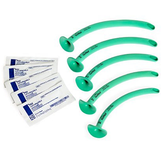
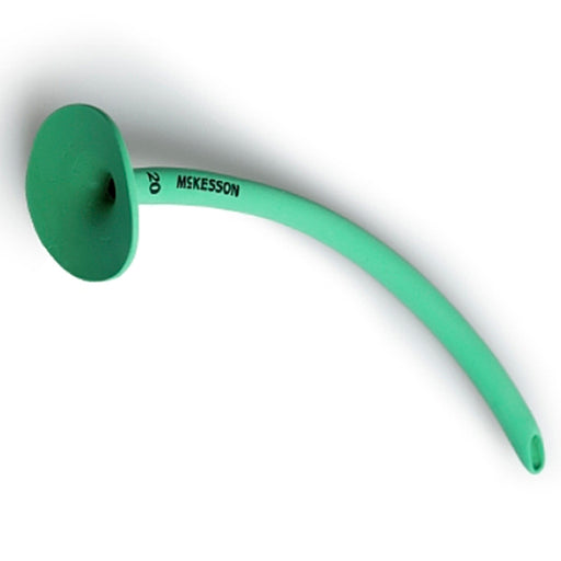
Nasopharyngeal Tube
-

 Sale 39%
Nasopharyngeal Airway Tube with Adjustable Flange by Rusch 28 French
Original price $ 17.95Current price $ 10.95 -
 Sale 28%
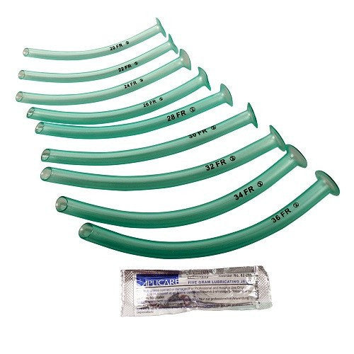
Nasopharyngeal Airway Set Trumpet Style (Robertazzi) with 28, 30, 32, 36 French Sizes Included
 Original price $ 89.95Current price $ 65.00
 -
 Sale 20%
Nasopharyngeal Airway Tubes, Robertazzi Style, 10/Box - McKesson
Original price $ 89.95Current price $ 72.00


Nasopharyngeal Tube (nasal) Airway Tube
A nasopharyngeal airway (NPA) – often called a “nasal trumpet” – is a hollow, flexible tube (usually plastic or soft rubber) inserted through the nose into the nasopharynx to maintain an open airway. It extends past the tongue and soft palate, keeping the airway patent by displacing the tongue and soft tissues that can block air flow. Unlike an oropharyngeal airway (which goes through the mouth), an NPA is placed nasally and generally does not trigger the gag reflex. This makes it useful for patients who are semi-conscious or have a strong gag reflex. In practice, a nasopharyngeal airway is used as a short-term adjunct to improve ventilation – for example, when bag-valve-mask ventilation alone is ineffective or when intubation is being prepared. It is a common emergency and anesthesia tool to “bypass” upper airway obstruction in a breathing patient until a more definitive airway (like endotracheal intubation) can be secured.
Indications (When and why it’s used)
Nasopharyngeal airways are indicated in patients who need help keeping their airway open but cannot tolerate an oropharyngeal airway. Typical uses include:
- Airway obstruction in a semi-conscious patient: If a patient is drowsy or intoxicated and the tongue or soft tissues are falling back to block the airway, an NPA can “beat” this obstruction. It is often used when bag-valve-mask ventilation fails to maintain adequate breathing and when intubation is not yet done.
- Intact gag reflex or clenched jaw: Because NPAs usually do not provoke vomiting or gagging, they are preferred over oral airways when the patient has an intact reflex or cannot open their mouth (for example, in seizure activity or jaw injury).
- Emergency airway adjunct: As a temporary measure in emergencies (trauma, resuscitation, airway compromise), an NPA quickly establishes an airway before definitive measures.
In summary, an NPA is a rescue airway device used to improve oxygenation and ventilation in spontaneously breathing patients who are at risk of airway obstruction (e.g. due to tongue fall-back), particularly when an oropharyngeal airway is inappropriate.
Contraindications and Precautions
Care must be taken with NPAs, as there are situations where they should not be used. Absolute contraindications include any suspicion of basal skull fracture or significant facial/nasal trauma – inserting the tube could injure the brain or circulatory structures. Similarly, patients with known midface fractures, nasal bone fractures, or severe head injuries should never receive an NPA. Relative contraindications (use with caution) include: recent nasal or sinus surgery, large nasal polyps, significant nasal septal deviation, coagulopathy or anticoagulation (because of bleeding risk), and severe sinusitis. In a nutshell, do not use a nasopharyngeal tube if there is any doubt about skull or facial integrity, or if the patient has acute nose pathology.
Potential complications of nasopharyngeal airway placement include:
- Nasal bleeding (epistaxis): Trauma to the nasal mucosa is common, so gentle insertion with lube is essential.
- Nasal or turbinate injury: The tube can fracture delicate nasal bones or turbinates if forced.
- Intracranial placement: In rare cases of unrecognized skull base fracture, the tube can enter the cranial vault (a catastrophic complication).
- Other issues: Improper sizing or placement can cause nasal tissue laceration, sinusitis over time, or even channel oxygen/air into the stomach if pushed too far.
In short, a nasopharyngeal (nasal) airway is a soft plastic/rubber tube inserted through the nose to keep the upper airway open. It is often called a “nasal trumpet” and is especially useful when an oral airway cannot be used (e.g. patient has a gag reflex or clenched jaw). When used correctly and sized/lubricated properly, it helps bypass upper airway obstruction by the tongue, improving ventilation in semi-conscious patients. Clinicians must select the correct length (usually measured from nostril to earlobe or angle of the jaw) and use water-based lubricant. It is a temporary measure – once applied, the patient should be closely monitored and planned for definitive airway management as needed. Always remember the contraindications (skull/facial trauma) and use caution due to bleeding risks