
Hypodermic Needles, Hypodermic Syringes for BD hypodermic needles


Hypodermic Needles
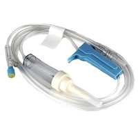
A hypodermic needle is a very thin, hollow metal tube with a sharp pointed bevel designed to pierce the skin and underlying tissues. It is typically made of stainless steel (or similar alloys) formed by tube drawing, and the distal tip is bevelled to create a sharp point that penetrates easily. Hypodermic needles attach to a syringe via a Luer-style hub (slip-fit or Luer-Lok), allowing precise control of injected or withdrawn fluids. In practice each needle is paired with a sterile syringe and is intended for single use: a hypodermic syringe can retain blood or medication residue indefinitely, so stringent practice dictates using a new sterile needle-syringe assembly for each injection to prevent contamination or infection.
Uses and Applications
Hypodermic needles are used wherever fluid must be introduced into or removed from the body under sterile conditions:
- Medication administration: Injecting drugs, vaccines or anesthetics into tissues. For example, intramuscular (IM) shots (e.g. vaccines, antibiotics) and subcutaneous (SC) injections (e.g. insulin) commonly use hypodermic needles.
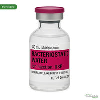
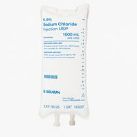
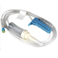
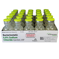
- Intravenous (IV) therapy: Accessing veins for fluid infusions or injections. Large-bore needles (low gauge) enable rapid fluid delivery in emergencies (e.g. trauma resuscitation) or are used to insert intravenous catheters.
- Blood collection/transfusion: Drawing blood samples or donating blood. In practice, a 21-gauge needle (green hub) is standard for routine phlebotomy, whereas larger needles (16–17G) are used for whole-blood donation or rapid transfusion because the wider bore allows high flow rates.
- Intradermal and pediatric injections: Very fine needles (high gauge) are used for delicate procedures. For example, intradermal tests (e.g. tuberculosis mantoux) or pediatric insulin injections use very fine needles (often 25–30G) to minimize pain.
- Laboratory and research use: Hypodermic needles can inoculate sterile media or inject reagents in lab animals/tissue cultures. Their smooth stainless-steel surface and fine bevel reduce contamination when injecting into agar plates or culture vials.
There are many injection routes: intravenous (into a vein) and intramuscular (into muscle) are among the most common; other routes include subcutaneous (into fat under the skin) and intradermal (into the skin layers). Hypodermic needles also see use in veterinary medicine, tattooing or body art (tattoo machines use pigment-filled needles), forensic sampling, and in consumer devices (e.g. insulin pens with attachable needles).
Injection Techniques and Handling
Proper technique ensures efficacy and safety:
- Needle selection: Choose needle gauge (thickness) and length appropriate to the patient and route. Thicker (lower-G) needles are used for deeper, high-flow needs (e.g. IM or IV), while thinner (higher-G) needles are used for small-volume or superficial injections. Typical lengths range from 3/8″ (for shallow infant injections) to 1″ or more (for large adults IM injections).
- Angle of insertion: For intramuscular injections, the needle is typically inserted at a 90° angle to the skin into the muscle (to reach deep tissue). For subcutaneous injections, a 45° angle is common (or even 90° with pinched skin if using a very short needle) to deposit medication just under the skin. Intradermal injections (e.g. allergy or TB tests) use a very shallow angle (~10–15°) to inject between skin layers. Intravenous injections are usually done at ~15–30° into a vein to allow a brief “flash” of blood return.
- Aseptic preparation: Clean the skin with an alcohol swab or antiseptic, let it dry, and wear gloves. Assemble the syringe-needle with sterile technique.
- Injection: Stretch or stabilize the skin (sometimes with the non-dominant hand or using the “Z-track” gluteal technique) to reduce movement. Quickly jab the needle to minimize pain (as recommended in best-practice guides). After insertion, some protocols (especially older IM technique) advise gently aspirating (pulling back on the syringe plunger) for 3–5 seconds to check for blood; if blood appears, the needle is in a vessel and should be withdrawn. (Note: current guidelines may or may not require routine aspiration depending on the injection site and medication.) Then depress the plunger steadily to inject the medication.
- Needle removal and care: Withdraw the needle quickly at the same angle of entry, apply gentle pressure or a bandage to the site if needed, and dispose of the needle-and-syringe immediately into a sharps container without recapping. Reusing or recapping needles is strongly discouraged to prevent needlestick injuries and contamination. Each injection must use a fresh, sterile needle and syringe.
- Patient comfort: Finer gauge needles and quick insert/removal minimize pain. For example, clinical guidelines note that 21G (green hub) or 23G (blue hub) needles are generally small enough to minimize tissue trauma yet large enough for most IM injections.
Needle Gauge and Color Coding
Hypodermic needles are specified by gauge (G), a numerical scale inversely related to diameter: a lower gauge number means a larger bore (wider needle), and vice versa. The standard measurement for needle gauge is the Birmingham wire gauge system (distinct from the French catheter scale). Needles range roughly from about 7G (very large, 4.5 mm OD) down to 34G (very fine, ~0.18 mm OD) in medical use. Common gauges for routine use lie between about 14G and 30G. For example, 21G–23G needles are used for intramuscular injections, whereas 25G–30G needles are used for insulin and pediatric injections. The choice depends on medication viscosity and required flow rate: thick fluids (like contrast dyes or whole blood) need low-gauge needles (16–20G), while watery solutions (like many vaccines or insulin) can be given through high-gauge needles (26G–30G).
To facilitate quick identification, needle hubs are color-coded by gauge according to ISO 6009 standards (though slight variations exist by manufacturer). Common gauge–color pairs include:
- 14G: Orange
- 16G: Grey
- 18G: Green
- 19G: White
- 20G: Pink
- 21G: Green
- 22G: Dark blue
- 23G: Blue
- 24G: Yellow
- 25G: Orange
- 26G: Violet
Finer needles beyond 26G have their own codes: for instance, 30G is typically purple. (Conversely, some even lower gauges like 12–14G are occasionally orange or grey, but these are mostly used in large-volume IV cannulas or biomed research, not routine injections.)
In summary, hypodermic needles come in many diameters and lengths. By choosing an appropriate gauge (color-coded on the hub) and following proper technique (site, angle, and aseptic handling) clinicians can safely deliver medications or draw fluids as needed.