Filters
- Blepharitis (1)
- Buy erythromycin (1)
- doctor-only (3)
- erythromycin 0.5% for babies (1)
- erythromycin eye ointment (1)
- Erythromycin near me (1)
- Erythromycin Ointment (1)
- Erythromycin Ophthalmic (1)
- erythromycin ophthalmic for infants (1)
- ewborn eye ointment (1)
- Eye antibiotic (3)
- eye infection prevention (1)
- Eye Infections (3)
- Keratitis (1)
- neonatal conjunctivitis (1)
- newborn dose (1)
- ophthalmia neonatorum prophylaxis (1)
- Pink Eye (3)
- Prevent Eye Infections (1)
- Antibiotic Eye Drops (1)
- Antibiotic Medication (1)
- Antibiotics (1)
- cipro eye drops (1)
- Ciprofloxacin (1)
- Ciprofloxacin Drops (1)
- ciprofloxacin eye (1)
- Ciprofloxacin Eye Drops (1)
- ciprofloxacin eye drops dosage (1)
- ciprofloxacin eye drops for pink eye (1)
- ciprofloxacin eye drops side effects (1)
- ciprofloxacin eye drops uses (1)
- ciprofloxacin ophthalmic (1)
- Conjunctivitis (1)
- Eye bacteria (1)
- Eye health (1)
- Fluoroquinolone Antibiotic (1)
- Bacteria In Eye (1)
- Ciloxan (1)
- Ciprofloxacin Ointment (1)
- Eye Bacteria (1)
- Ophthalmic Ointment (1)
- Treat Bacteria in Eye (1)
- Treat bacterial infections (1)
Ophthalmic Supplies, instruments & Equipment to Eye antibiotic
Ophthalmic Supplies, Instruments & Equipment
Ophthalmic (eye‐care) supplies encompass all the tools and devices used to examine, diagnose and treat eye diseases and vision problems. This includes diagnostic instruments for testing and imaging the eye, surgical instruments for eye operations, andConsumable supplies used in routine eye care. For example, instruments like ophthalmoscopes (to view the retina), retinoscopes (for measuring refractive error), keratometers (to measure corneal curvature) and slit-lamp microscopes (for detailed anterior eye exams) are standard ophthalmic tools. A typical eye exam also uses a phoropter or trial lens set to determine a patient’s prescription by having them look through different lenses (see image below), and a tonometer to measure intraocular pressure (screening for glaucoma).
-
Vision testing equipment: Visual acuity charts (Snellen charts) and occluders test distance vision. Automated autorefractors/keratometers estimate the eye’s prescription and corneal shape. The clinician then refines the prescription with a phoropter or trial-frame (as shown) and retinoscope. An ophthalmoscope (handheld or slit-lamp/indirect) is used to inspect the retina and optic nerve. (As a reference summary notes, these specialized instruments let doctors “study a patient’s eyes and prescribe spectacles,” including devices to observe the retina and measure refractive error.)
-
Slit lamp biomicroscope: A high-intensity microscope with an adjustable slit beam (illumination) is used to examine the front of the eye – cornea, iris, lens and anterior chamber – in great detail. It often uses special lenses to also view the retina through the pupil. This is a cornerstone of the eye exam for detecting cataracts, corneal scars or infections and tracking diseases like macular degeneration.
-
Tonometer (eye pressure instruments): Devices such as Goldmann applanation tonometers, non-contact “puff” tonometers, or handheld tonopen measure intraocular pressure. Elevated pressure is a key glaucoma risk, so tonometry is routinely performed in any eye exam.
-
Visual field analyzers: Perimetry machines (e.g. Humphrey Field Analyzer) map a patient’s peripheral (side) vision. This is important for detecting blind spots from glaucoma, retinal disease or neurological problems affecting the visual pathways.
-
Imaging equipment: Specialized cameras and instruments capture images of internal structures. Fundus cameras photograph the retina and optic nerve, often with dye (fluorescein) for angiography. Optical Coherence Tomography (OCT) uses laser light to create cross-sectional images of the retina and optic nerve, crucial in diagnosing macular degeneration or glaucoma. Ultrasonic A/B-scan devices measure eyeball length (A-scan) or image posterior segments through opaque media (B-scan) in cases of dense cataract or vitreous hemorrhage.
-
Surgical instruments: For eye surgery (cataract, glaucoma, retina, oculoplastics), specialized microsurgical tools are used. These include eye specula (to hold eyelids open), delicate forceps and scissors (e.g. Vannas scissors, capsulorhexis forceps), and fine needles for suturing. A phacoemulsification machine uses ultrasonics to break up and aspirate a cloudy lens (cataract). An operating microscope provides high magnification for precision. Lasers (Nd:YAG, argon, excimer) are common – e.g. YAG lasers clear posterior capsule opacities after cataract surgery or perform laser trabeculoplasty for glaucoma. Cryoprobes, vitrectors, and injectors for intraocular lenses are also used.
-
Supportive supplies: These include all consumables needed for exams and procedures. Eye drops/medications (topical anesthetics, dilating drops, antibiotic or anti-inflammatory drops) are essential. Sterile items like gauze, sterile saline for irrigation, drapes, and surgical gloves protect the eye and maintain asepsis. Eye patches/shields are used post-procedure or for amblyopia therapy. Ophthalmic measurements and low-vision aids – trial lens sets, prisms, Maddox rods, occluders – support vision testing and therapy. Even eyeglasses/contact lens supplies (like lensometers and solutions) are often part of an ophthalmic office.
In summary, ophthalmic equipment covers everything needed for a full eye exam and eye care: from refraction tools and microscopes for diagnosis, to cameras and visual field machines for imaging, to lasers and microsurgical instruments for treatment. Each item has a specific role – for instance, a phoropter precisely adjusts lens power for refractive testing (as in the photo above), while a tonometer measures intraocular pressure to check for glaucoma. Authoritative references note that these “specialized instruments” let eye doctors study a patient’s eye (retina, cornea, lens) and determine refractive errors. Together, they enable comprehensive vision testing, disease detection (cataract, glaucoma, retinal disease) and safe eye surgery.
-
 Sale 44%
Original price $ 29.95Current price $ 16.89
Sale 44%
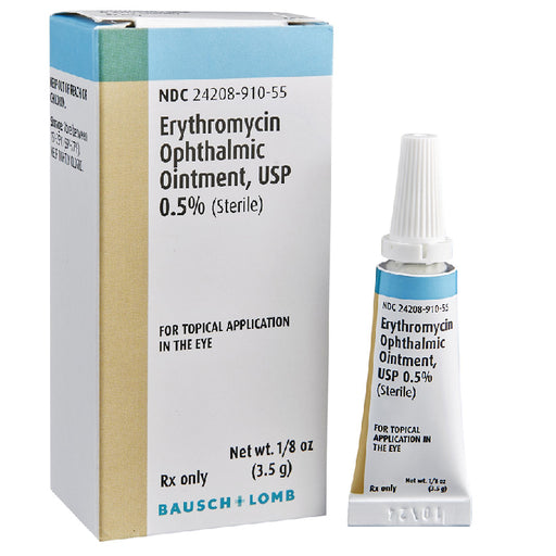
Original price $ 29.95Current price $ 16.89Erythromycin Eye Ointment 0.5%, Sterile 3.5 grams (Rx)
2 reviewsErythromycin Ophthalmic Ointment 0.5% is the leading FDA-approved, prescription antibiotic ointment for the treatment and prevention of a wide rang...
View full details🔒 Medical License Required -
 Sale 35%
Original price $ 22.95Current price $ 14.95
Sale 35%
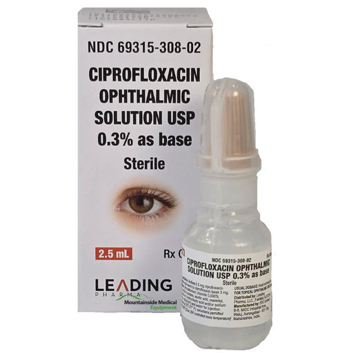
Original price $ 22.95Current price $ 14.95Ciprofloxacin Eye Drops 0.3% 2.5 mL (RX)
Ciprofloxacin Eye Drops Ciprofloxacin eye drops (0.3% ophthalmic solution; brand name Ciloxan) are a broad-spectrum fluoroquinolone antibiotic used...
View full details🔒 Medical License Required -
 Sale 10%
Original price $ 399.95Current price $ 359.00
Sale 10%
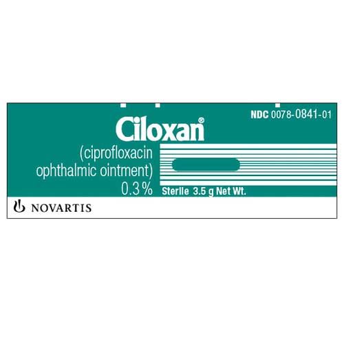
Original price $ 399.95Current price $ 359.00Ciloxan Ciprofloxacin Ophthalmic Ointment 0.3%. Sterile 3.5 gram (RX)
Ciloxan Ciprofloxacin Ointment is a topical antibiotic medication used to treat infections of the eye, specifically conjunctivitis (pink eye) cause...
View full details🔒 Medical License Required


Ophthalmic Supplies, Instruments & Equipment
Ophthalmic (eye‐care) supplies encompass all the tools and devices used to examine, diagnose and treat eye diseases and vision problems. This includes diagnostic instruments for testing and imaging the eye, surgical instruments for eye operations, andConsumable supplies used in routine eye care. For example, instruments like ophthalmoscopes (to view the retina), retinoscopes (for measuring refractive error), keratometers (to measure corneal curvature) and slit-lamp microscopes (for detailed anterior eye exams) are standard ophthalmic tools. A typical eye exam also uses a phoropter or trial lens set to determine a patient’s prescription by having them look through different lenses (see image below), and a tonometer to measure intraocular pressure (screening for glaucoma).
-
Vision testing equipment: Visual acuity charts (Snellen charts) and occluders test distance vision. Automated autorefractors/keratometers estimate the eye’s prescription and corneal shape. The clinician then refines the prescription with a phoropter or trial-frame (as shown) and retinoscope. An ophthalmoscope (handheld or slit-lamp/indirect) is used to inspect the retina and optic nerve. (As a reference summary notes, these specialized instruments let doctors “study a patient’s eyes and prescribe spectacles,” including devices to observe the retina and measure refractive error.)
-
Slit lamp biomicroscope: A high-intensity microscope with an adjustable slit beam (illumination) is used to examine the front of the eye – cornea, iris, lens and anterior chamber – in great detail. It often uses special lenses to also view the retina through the pupil. This is a cornerstone of the eye exam for detecting cataracts, corneal scars or infections and tracking diseases like macular degeneration.
-
Tonometer (eye pressure instruments): Devices such as Goldmann applanation tonometers, non-contact “puff” tonometers, or handheld tonopen measure intraocular pressure. Elevated pressure is a key glaucoma risk, so tonometry is routinely performed in any eye exam.
-
Visual field analyzers: Perimetry machines (e.g. Humphrey Field Analyzer) map a patient’s peripheral (side) vision. This is important for detecting blind spots from glaucoma, retinal disease or neurological problems affecting the visual pathways.
-
Imaging equipment: Specialized cameras and instruments capture images of internal structures. Fundus cameras photograph the retina and optic nerve, often with dye (fluorescein) for angiography. Optical Coherence Tomography (OCT) uses laser light to create cross-sectional images of the retina and optic nerve, crucial in diagnosing macular degeneration or glaucoma. Ultrasonic A/B-scan devices measure eyeball length (A-scan) or image posterior segments through opaque media (B-scan) in cases of dense cataract or vitreous hemorrhage.
-
Surgical instruments: For eye surgery (cataract, glaucoma, retina, oculoplastics), specialized microsurgical tools are used. These include eye specula (to hold eyelids open), delicate forceps and scissors (e.g. Vannas scissors, capsulorhexis forceps), and fine needles for suturing. A phacoemulsification machine uses ultrasonics to break up and aspirate a cloudy lens (cataract). An operating microscope provides high magnification for precision. Lasers (Nd:YAG, argon, excimer) are common – e.g. YAG lasers clear posterior capsule opacities after cataract surgery or perform laser trabeculoplasty for glaucoma. Cryoprobes, vitrectors, and injectors for intraocular lenses are also used.
-
Supportive supplies: These include all consumables needed for exams and procedures. Eye drops/medications (topical anesthetics, dilating drops, antibiotic or anti-inflammatory drops) are essential. Sterile items like gauze, sterile saline for irrigation, drapes, and surgical gloves protect the eye and maintain asepsis. Eye patches/shields are used post-procedure or for amblyopia therapy. Ophthalmic measurements and low-vision aids – trial lens sets, prisms, Maddox rods, occluders – support vision testing and therapy. Even eyeglasses/contact lens supplies (like lensometers and solutions) are often part of an ophthalmic office.
In summary, ophthalmic equipment covers everything needed for a full eye exam and eye care: from refraction tools and microscopes for diagnosis, to cameras and visual field machines for imaging, to lasers and microsurgical instruments for treatment. Each item has a specific role – for instance, a phoropter precisely adjusts lens power for refractive testing (as in the photo above), while a tonometer measures intraocular pressure to check for glaucoma. Authoritative references note that these “specialized instruments” let eye doctors study a patient’s eye (retina, cornea, lens) and determine refractive errors. Together, they enable comprehensive vision testing, disease detection (cataract, glaucoma, retinal disease) and safe eye surgery.